43 label skin layers
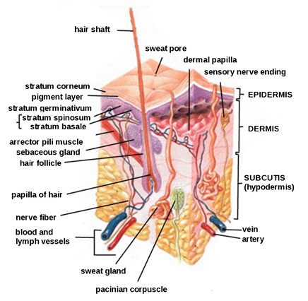
Understanding The Different Layers Of Skin - SkinKraft The three main layers in it are: Epidermis Dermis Hypodermis Functions Of The Skin's Layers 1. Epidermis Epidermis is the outermost layer of your skin, making it the protective barrier which prevents the entry of harmful bacteria, viruses and other foreign substances into the deeper layers. Layers of Skin: How Many, Diagram, Model, Anatomy, In Order The epidermis is the top layer of your skin. It's the only layer that is visible to the eyes. The epidermis is thicker than you might expect and has five sublayers. Your epidermis is constantly...
Skin Lesson for Kids: Layers & Parts | Study.com The hypodermis is the bottom most layer of skin. Just so you know, some people call this layer 'subcutaneous tissue' or 'subcutis'. The hypodermis is made mostly of fat. This fat helps to regulate ...
Label skin layers
Skin: Cells, layers and histological features | Kenhub This article will describe the anatomy and histology of the skin. Undoubtedly, the skin is the largest organ in the human body; literally covering you from head to toe. The organ constitutes almost 8-20% of body mass and has a surface area of approximately 1.6 to 1.8 m2, in an adult. It is comprised of three major layers: epidermis, dermis and ... Skin Diagram with Detailed Illustrations and Clear Labels Skin Diagram. The largest organ in the human body is the skin, covering a total area of about 1.8 square meters. The skin is tasked with protecting our body from the external elements as well as microbes. The skin is also responsible for maintaining our body temperature - this was apparent in victims who were subjected to the medival torture ... Skin: The Histology Guide - University of Leeds Some facts about skin. Skin is the largest organ of the body. It has an area of 2 square metres (22 square feet) in adults, and weighs about 5 kilograms. The thickness of skin varies from 0.5mm thick on the eyelids to 4.0mm thick on the heels of your feet. Skin is the major barrier between the inside and outside of your body!
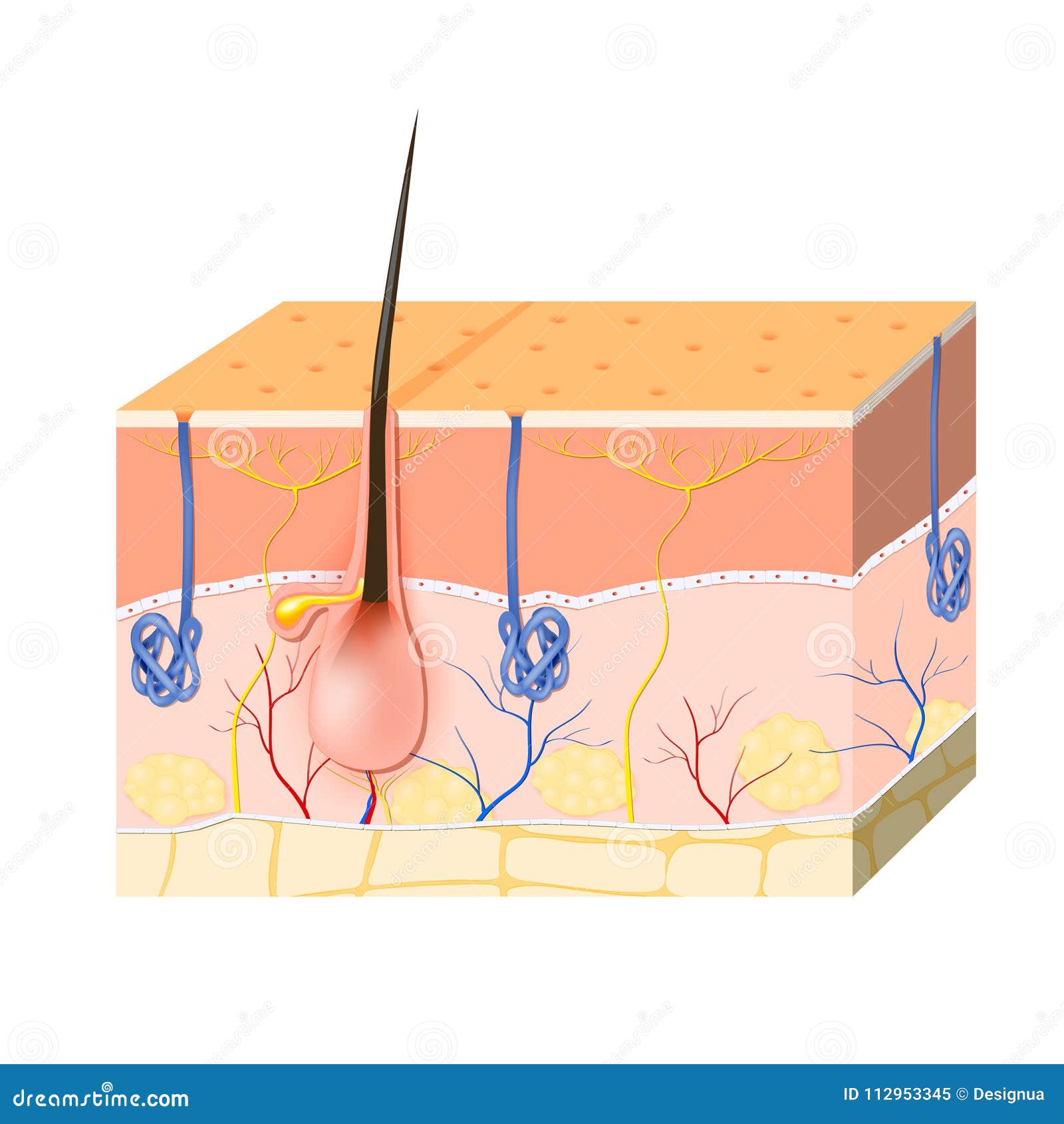
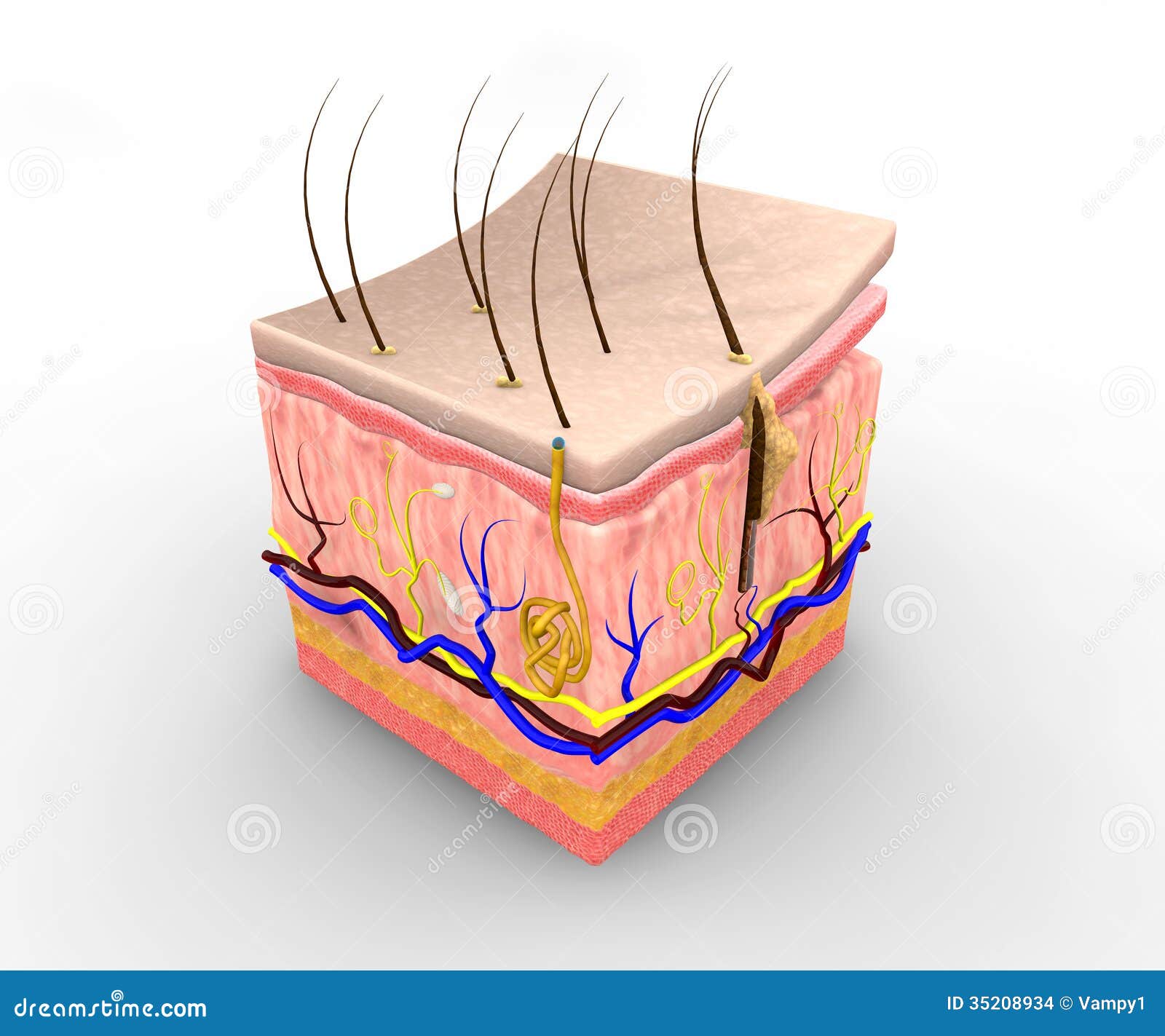
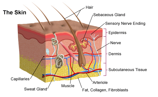
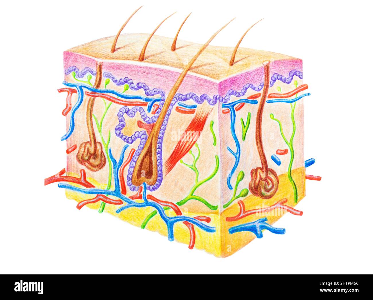
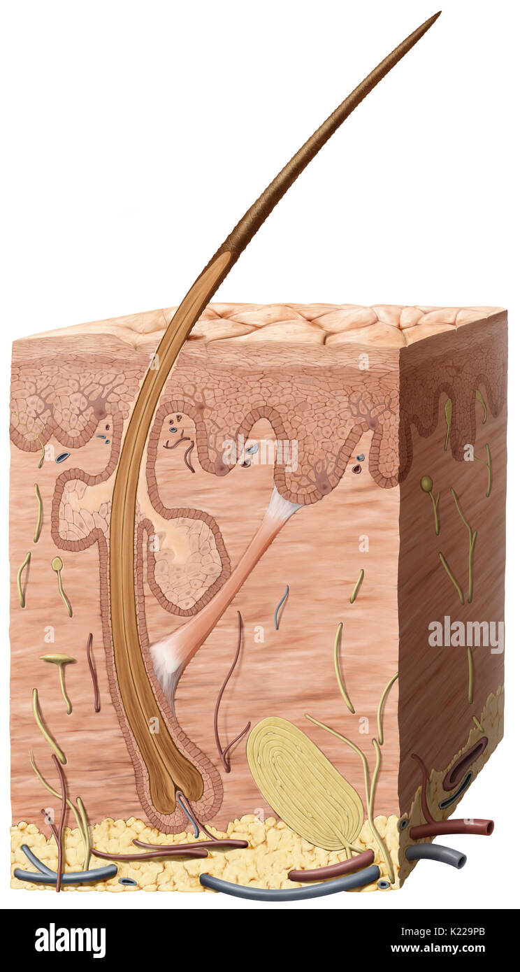
Label skin layers. diagram of label human skin Layers Of Skin - YouTube . skin layers. Hair Structure, Diagrams And Function Of The Hair Shaft Layers . anatomy follicle cosmetology hairdressing integumentary pancreas barbering. Under The Micrsocope: Onion Cell (100x - 400x) - YouTube . onion cell under 400x 100x Skin Anatomy: The Layers of Skin and Their Functions How many layers of skin are there? There are three main layers of skin: Epidermis: The outermost layer, which contains five sub-layers Dermis: The middle layer, which consists of two parts known as the papillary dermis (thin, upper layer) and the reticular dermis (thick, lower layer) Subcutaneous tissue: The deepest layer of skin Layers of the skin: label Diagram | Quizlet muscle layer Epidermis Dermis Hypodermis (Subcutaneous) Sweat pore Sebaceous gland Hair shaft Arrector pili muscle Pulls on the hair follicle to make it stand up (goose bumps) Papillae--part of the papillary layer Increases the surface area between the epidermis and dermis, providing oxygen and nutrients to the outermost layer Nerve cells The Skin (Human Anatomy): Picture, Definition, Function, and Skin ... The skin protects us from microbes and the elements, helps regulate body temperature, and permits the sensations of touch, heat, and cold. Skin has three layers: The epidermis, the outermost layer ...
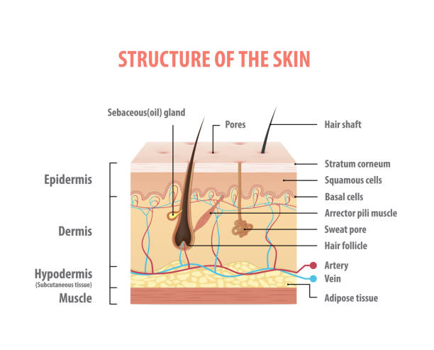
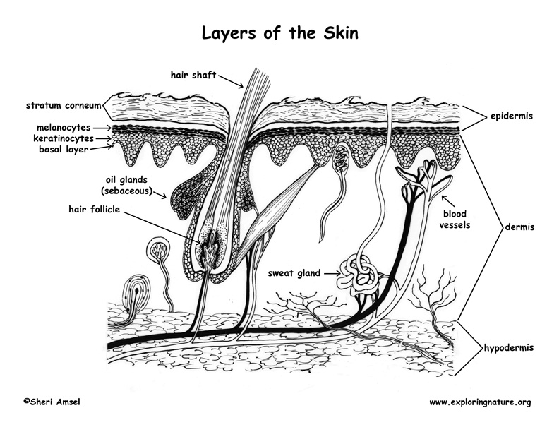
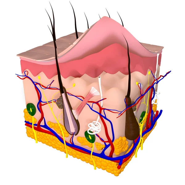
Layers Of Skin Tissue And Structure Here are a number of highest rated Layers Of Skin Tissue And Structure pictures upon internet. We identified it from well-behaved source. Its submitted by government in the best field. We consent this nice of Layers Of Skin Tissue And Structure graphic could possibly be the most trending topic once we part it in google help or facebook. The Skin: 7 Most Important Layers, Functions & Thickness It is made up of seven layers ( [starting from the top layer down to the bottom [deepest] layer): Stratum corneum Stratum lucidum Stratum granulosum Stratum spinosum Stratum basale Dermis Hypodermis The first five layers form the epidermis, which is the outermost, thick layer of the skin. Human Biology fig. 1.24 - Layers of the skin - English labels Layers of the skin. The inner layer of the skin is the dermis, and the outer layer is the epidermis. The epidermis can be specified further in the stratum corneum, stratum lucidum, stratum gransulosum, stratum spinosum and stratum basale. English labels. From 'Human Biology' by D. Wilkin and J. Brainard . Anatomical structures in item: Dermis A Human Body Skin-structure Quiz! - ProProfs Quiz Label F is: A. Reticular Layer of Dermis B. Dermal Papillae Layer C. Sensory Nerve Fiber D. Artery 8. Label G is: A. Sebaceous (oil) Gland B. Eccrine Sweat Gland C. Eccrine Sweat Gland D. Adipose Tissue 9. Label H is: A. Pacinian Corpuscle B. Arrector Pili Muscle C. Hair Follicle Receptor D. Pore 10. Label I is: A. Sensory Nerve Fiber B.
Label the diagram of the skin - Labelled diagram - Wordwall Hair shaft, Hair follicle, Arrector pili muscle, Sweat pore, Sebaceous gland, Sweat gland, Blood capillaries, Dermal Papila, Nerve endings, Epidermis, Dermis, Subcutaneous layer. Label the diagram of the skin Share by Charlotte98 Biology Like Edit Content More Leaderboard Log in required Theme Switch template Interactives Layers of the skin Labeling (Final Version) Diagram - Quizlet FIRST LAYER exposed surface of the skin consists of dead cells 15 to 30 layers of protective dead layers that are water resistant. changes fully every 25- 45 days stratum basale Attaches to the basal lamina (LAST LAYER) contains melanocytes, basal cells and Merkel cells. basal lamina Basement layer of the epidermis. sebaceous gland (oil gland) Anatomy, Skin (Integument), Epidermis - StatPearls - NCBI Bookshelf Skin is the largest organ in the body and covers the body's entire external surface. It is made up of three layers, the epidermis, dermis, and the hypodermis, all three of which vary significantly in their anatomy and function. The skin's structure is made up of an intricate network which serves as the body's initial barrier against pathogens, UV light, and chemicals, and mechanical injury. PDF Skin Diagram Labeling - New Providence School District Skin Diagram Labeling . 1. Label the diagram with the . ... You may label with a line or put the label directly onto the area described. Be as precise as possible. If you are worried about the precision of your label add a word ... U. Layers of keratinized cells V. Releases sebum . 2. Color the following structure on the diagram .
Ready & Popular Non-medical label for disposable packaging carton di ZiloongStore | Tokopedia
Label the skin - Teaching resources - Wordwall label the dialogue Labelled diagram. by Nessafowler. Appendages and layers of the skin Match up. by Sowens. The Skin diagram (black and white) Labelled diagram. by Jordannao. Label the Heart diagram Labelled diagram. by Rtomlin. Label the Skeleton Labelled diagram.
Skin Layers with Sebaceous Gland and Sweat Glands Stock ...
Skin Labeling | Biology Game | Turtle Diary Identify and label figures in Turtle Diary's interactive online game, Skin Labeling! Drag the given words to the correct blanks to complete the labeling! Upgrade to remove ads
Skin Layers Illustration 35208934 - Megapixl
Skin Labeling Quiz - PurposeGames.com This is an online quiz called Skin Labeling. There is a printable worksheet available for download here so you can take the quiz with pen and paper. From the quiz author. Epidermis, Dermis, Hypodermis ... Cell and Layers of Epidermis 14p Image Quiz. Playlists by same creator. marthamae's favorites 2 games. PurposeGames Create. Play. Learn.

The cells of the dermis become the epidermis | Skin anatomy ...
Label The Diagram Of The Layers Of The Skin - Learny Kids Displaying top 8 worksheets found for - Label The Diagram Of The Layers Of The Skin. Some of the worksheets for this concept are Integumentary system labeling work answers, Title skin structure, Integumentary system work basic skin structure, Label the skin anatomy diagram answers, Name your skin, Section through skin, Inside earth work, Anatomy physiology.

Unit 6: HBS integumentary system due 01/20/15
Label the layers of the skin Quiz - By mrumph - Sporcle Label the layers of the skin Can you name the Label the layers of the skin? By mrumph. Plays-/5-RATE QUIZ. YOU. MORE INFO Map. Forced Order Answers have to be entered in order Answers have to be entered in order hide this ad. PLAY QUIZ : % % Score. 0/4. Timer. 10:00. Give Up ...
Integumentary system review key
Skin Diagram Teaching Resources | Teachers Pay Teachers Test your kids/students' knowledge of human anatomy and the different parts of the human skin with this Parts of the Skin Diagram Worksheet.TAGS:Human Skin Labelling Worksheet, Label the Skin, Label the parts of the Skin, Skin Diagram to Color, Human Skin Anatomy Activity.PLEASE CHECK OUT THESE OTHER RESOURCES IN MY STORE:Animals Tracing and ...
Label the Layers of Skin by Little Learning Lane | TpT
PDF Title: Skin Structure - Kent State University Step 1 Ask students to identify the primary function of skin (protection). Ask students to identify any parts of the skin that they know. If they do not identify any, prompt them with the some key terms such as hair, follicle, blood vessel, and sweat gland.
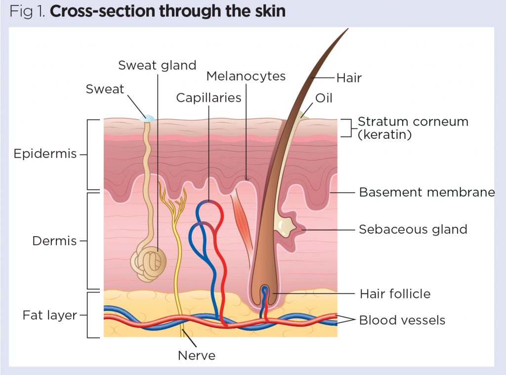
Skin 1: the structure and functions of the skin | Nursing Times
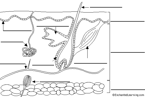
Label Skin Diagram Printout - EnchantedLearning.com dermis - (also called the cutis) the layer of the skin just beneath the epidermis. epidermis - the outer layer of the skin. hair follicle - a tube-shaped sheath that surrounds the part of the hair that is under the skin. It is located in the epidermis and the dermis.

Human Skin Image & Photo (Free Trial) | Bigstock
Solved Label the layers of the skin | Chegg.com Who are the experts? Experts are tested by Chegg as specialists in their subject area. We review their content and use your feedback to keep the quality high. 100% (38 ratings) Following are the labels of the skin from top to the bot …. View the full answer. Transcribed image text: Label the layers of the skin.
3,908 Skin Diagram Stock Photos, Pictures & Royalty-Free ...
The Three Layers of Skin and Their Functions | FLDSCC 3. The Hypodermis. The hypodermis is made of subcutaneous (under the skin) fats, connective tissues, blood vessels, and nerve cells. It's the layer of skin where fat is deposited and stored. The blood vessels in the hypodermis are bigger and connect to the rest of your body. Stored fat helps regulate body tissue and cushion your body's ...
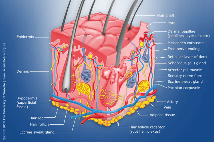
Diagram of human skin structure — Science Learning Hub
Label the Skin Layers - Printable - PurposeGames.com This is a free printable worksheet in PDF format and holds a printable version of the quiz Label the Skin Layers. By printing out this quiz and taking it with pen and paper creates for a good variation to only playing it online. This printable worksheet of Label the Skin Layers is tagged.
Label Skin Diagram Printout - EnchantedLearning.com
Skin: The Histology Guide - University of Leeds Some facts about skin. Skin is the largest organ of the body. It has an area of 2 square metres (22 square feet) in adults, and weighs about 5 kilograms. The thickness of skin varies from 0.5mm thick on the eyelids to 4.0mm thick on the heels of your feet. Skin is the major barrier between the inside and outside of your body!
Skin: Cells, layers and histological features | Kenhub
Skin Diagram with Detailed Illustrations and Clear Labels Skin Diagram. The largest organ in the human body is the skin, covering a total area of about 1.8 square meters. The skin is tasked with protecting our body from the external elements as well as microbes. The skin is also responsible for maintaining our body temperature - this was apparent in victims who were subjected to the medival torture ...

Raul Blog: Unlabeled Skin Layer Diagram
Skin: Cells, layers and histological features | Kenhub This article will describe the anatomy and histology of the skin. Undoubtedly, the skin is the largest organ in the human body; literally covering you from head to toe. The organ constitutes almost 8-20% of body mass and has a surface area of approximately 1.6 to 1.8 m2, in an adult. It is comprised of three major layers: epidermis, dermis and ...

Chapter 6 - Labeling Parts of Skin - sweat gland blood ...
Integumentary system parts: Quizzes and diagrams | Kenhub

Anatomy 3.2 Integumentary System: Epidermis Labeling Diagram ...

Layers of Skin: How Many, Diagram, Model, Anatomy, In Order

205 Wk1 Label the skin layers Quiz - By TraceyH100
Skin - Teaching resources

5.1 Layers of the Skin – Anatomy & Physiology
Anatomy of the Skin

21,424 Skin Anatomy Stock Photos, Pictures & Royalty-Free ...

91 Skin Diagram To Label Illustrations & Clip Art - iStock

Human skin - Wikipedia
Skin layers hi-res stock photography and images - Alamy

Human Skin Anatomy Cross Section Diagram Stock Illustration ...

Skin Layers Teaching Resources | Teachers Pay Teachers

Skin Labeling Quiz
Layers of the Skin

About Melanoma - Dana-Farber Cancer Institute | Boston, MA
Human skin layers Stock Photos, Royalty Free Human skin ...

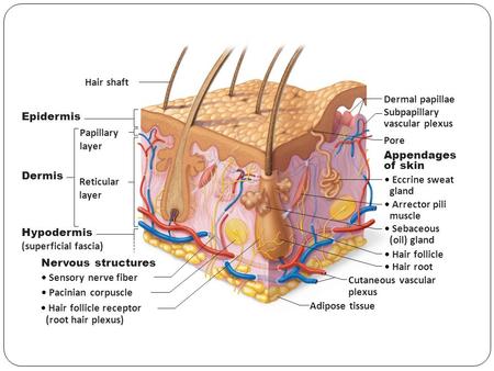
Schematic representation of the skin layers and appendage ...

36,269 Epidermis Images, Stock Photos & Vectors | Shutterstock

File:Skin Layers Unlabeled.jpg - Wikimedia Commons

File:Labeled layers of the skin.jpg - Wikimedia Commons

Skin Diagram with Detailed Illustrations and Clear Labels
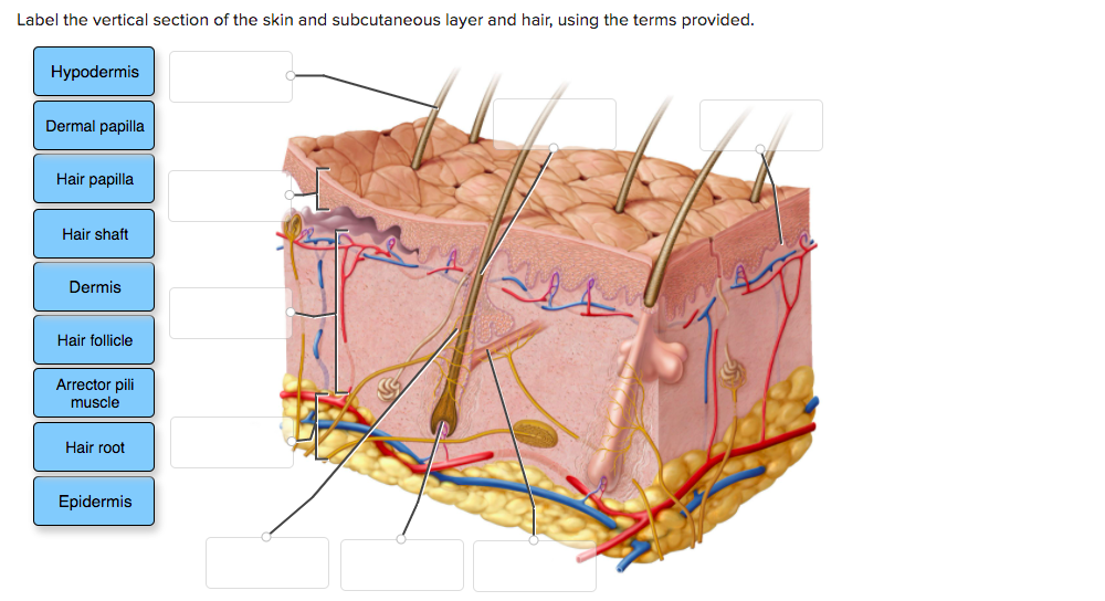
Solved Label the vertical section of the skin and | Chegg.com

Draw in easy steps: How to draw skin LS
Also known as… The SKIN! EPIDERMIS Pages ppt download
6.5: Laboratory Activities and Assignment - Biology LibreTexts
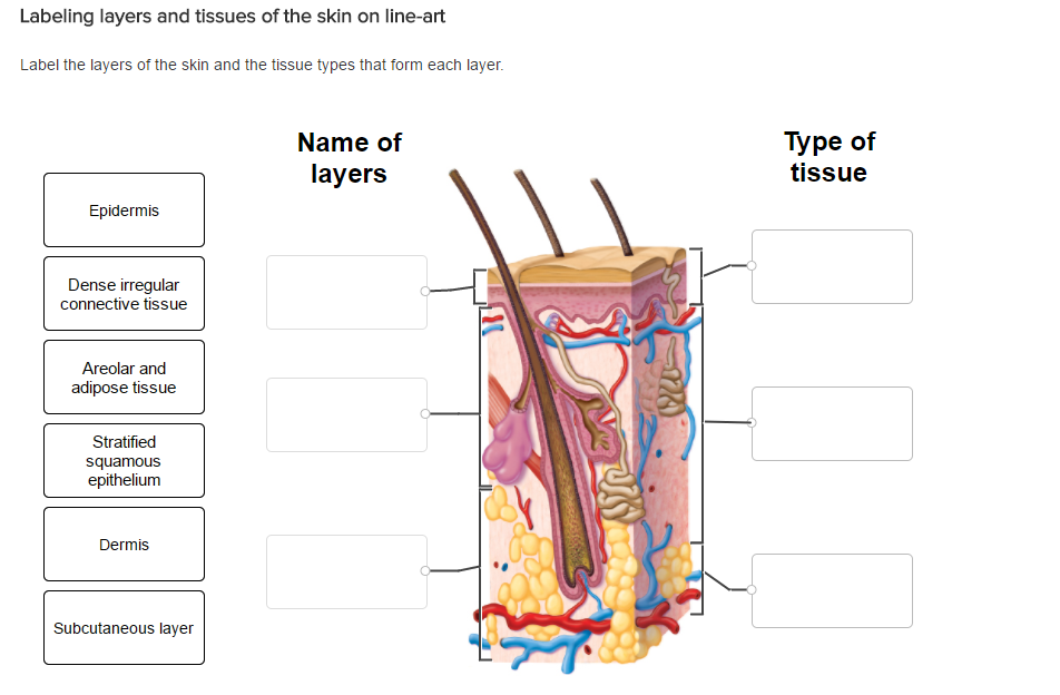
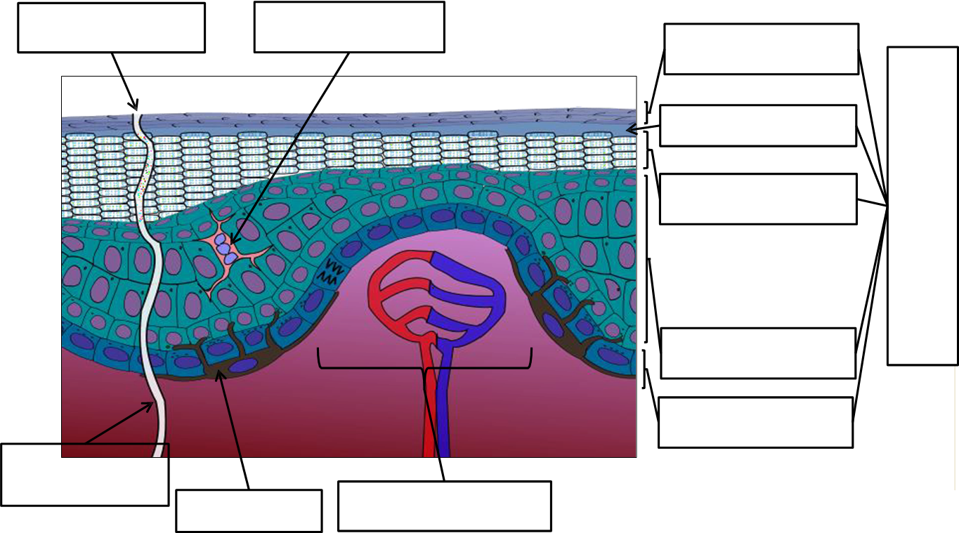
Solved Label the layers of the skin and the tissue types ...
Human skin cross section hi-res stock photography and images ...
Post a Comment for "43 label skin layers"