42 transmission electron micrograph labeled
› topics › agricultural-andTransmission Electron Microscopy - an overview ... 3.325.5.6 Transmission Electron Microscopy. TEM provides high-resolution imaging and is used for studying small areas or even single mineral platelets selectively. The TEM can be operated at different electron energies (often 100 keV for conventional TEM and 1 MeV for high-resolution imaging). The contrast of the TEM images is dependent on ... Ebolavirus - Wikipedia WebThe genus Ebolavirus (/ i ˈ b oʊ l ə / - or / ə ˈ b oʊ l ə ˌ v aɪ r ə s /; ee-BOH-lə- or ə-BOH-lə-VY-rəs) is a virological taxon included in the family Filoviridae (filament-shaped viruses), order Mononegavirales. The members of this genus are called ebolaviruses, and encode their genome in the form of single-stranded negative-sense RNA. The six known virus …
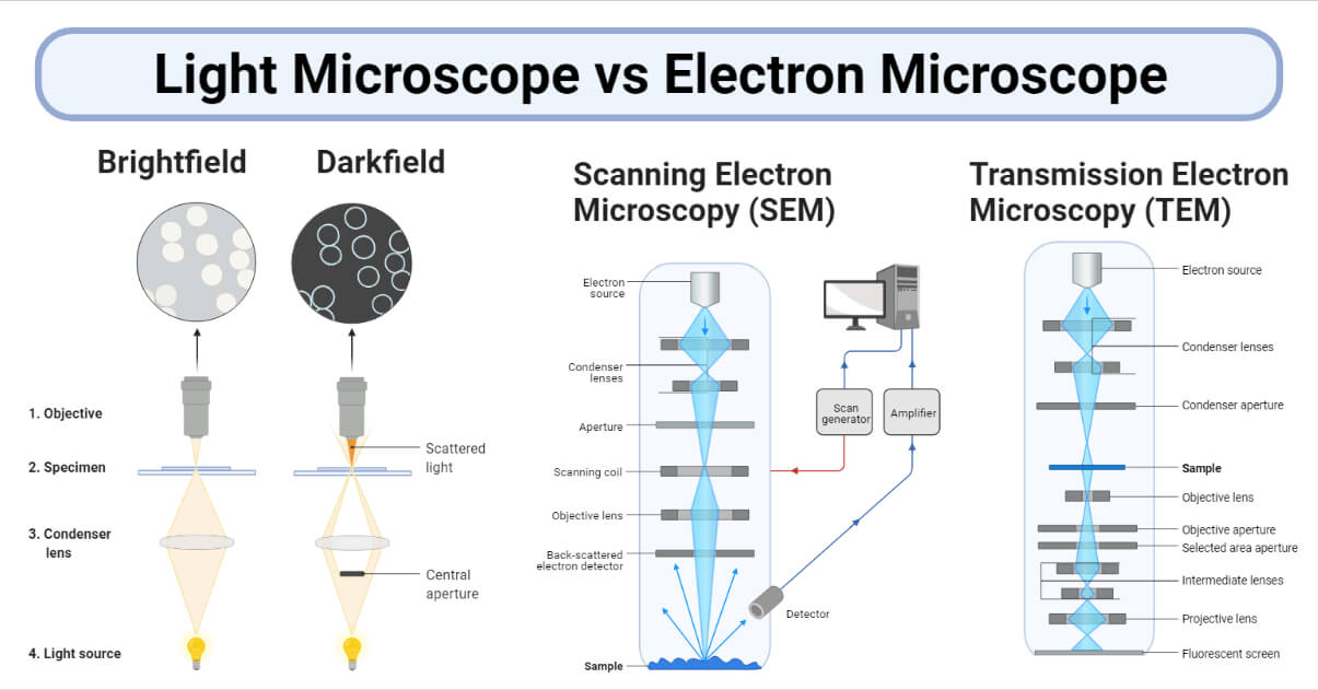
Electron Microscope Principle, Uses, Types and Images (Labeled ... Oct 3, 2022 ... It is called a scanning electron microscope because the image is formed by scanning the surface of the specimen in a raster pattern using a ...
Transmission electron micrograph labeled
Transmission electron microscopy - Wikipedia Transmission electron microscopy (TEM) is a microscopy technique in which a beam of electrons is transmitted through a specimen to form an image. The Transmission Electron Microscope | CCBER TEMs employ a high voltage electron beam in order to create an image. An electron gun at the top of a TEM emits electrons that travel through the microscope's ... Virtual EM Micrograph List | histology - University of Michigan WebFat Cells: This electron micrograph depicts mature fat cells. You can see one large lipid droplet in the cytoplasm of each cell. The nuclei of many cells are not included in the field of view. Brown fat cells would have several small lipid droplets all of which would be roughly the same size. Remember that each fat cell is enclosed by a thin basal lamina …
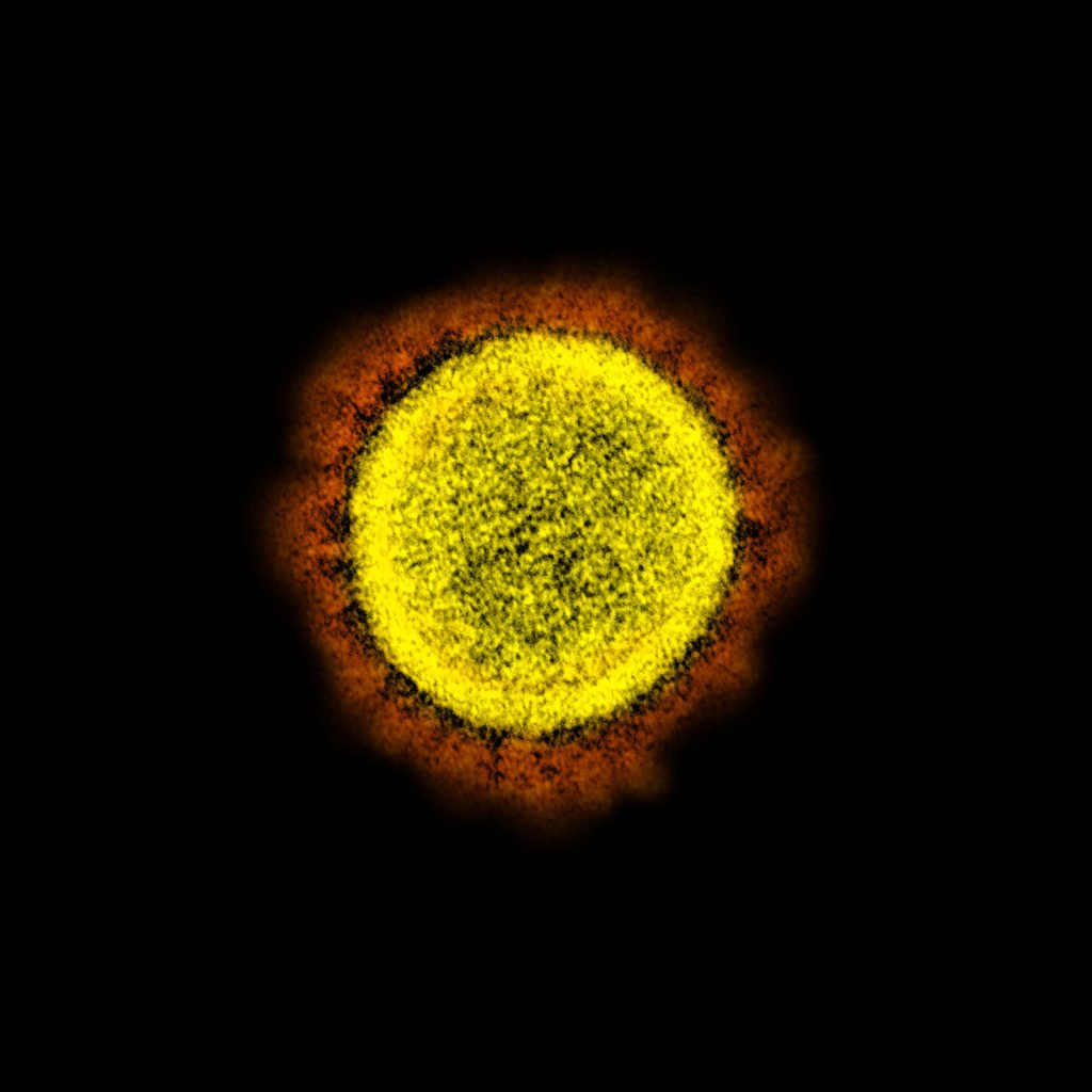
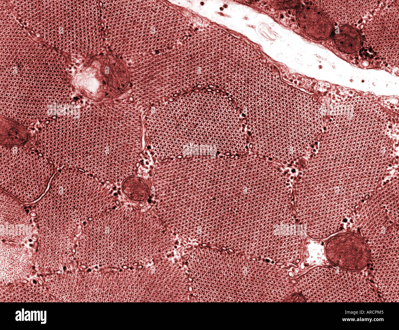
Transmission electron micrograph labeled. Processing tissue and cells for transmission electron microscopy in ... Oct 4, 2007 ... In transmission electron microscopy (TEM), electrons are transmitted through a plastic-embedded specimen, and an image is formed. 1.2 Skill: Interpretation of electron micrographs - YouTube Dec 25, 2015 ... Interpretation of electron micrographs to identify organelles and deduce the functions of specialized cells. Electron micrographs used with ... Smallpox - Wikipedia WebThis transmission electron micrograph depicts a number of smallpox virions. The "dumbbell-shaped" structure inside the virion is the viral core, which contains the viral DNA; Mag. = ~370,000× Virus classification (unranked): Virus: Realm: Varidnaviria: Kingdom: Bamfordvirae: Phylum: Nucleocytoviricota: Class: Pokkesviricetes: Order: Chitovirales: … › figure › TransmissionTransmission electron micrograph of a de-epithelialised ... Download scientific diagram | Transmission electron micrograph of a de-epithelialised gastric mucosal sample cultured for 24 hours. Discrete pores (P) in the basement membrane are in continuity ...
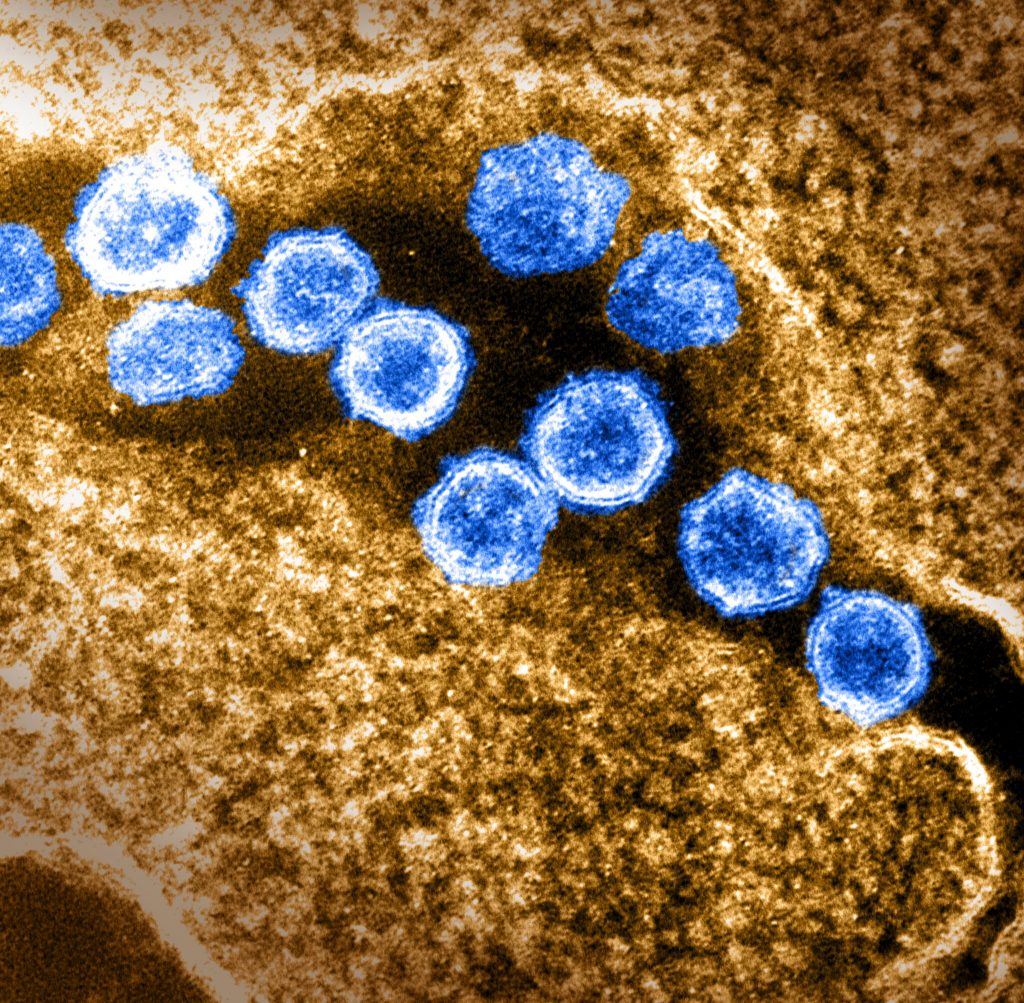
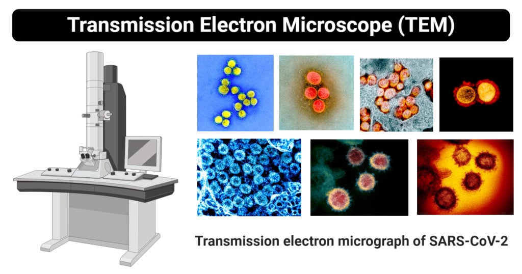
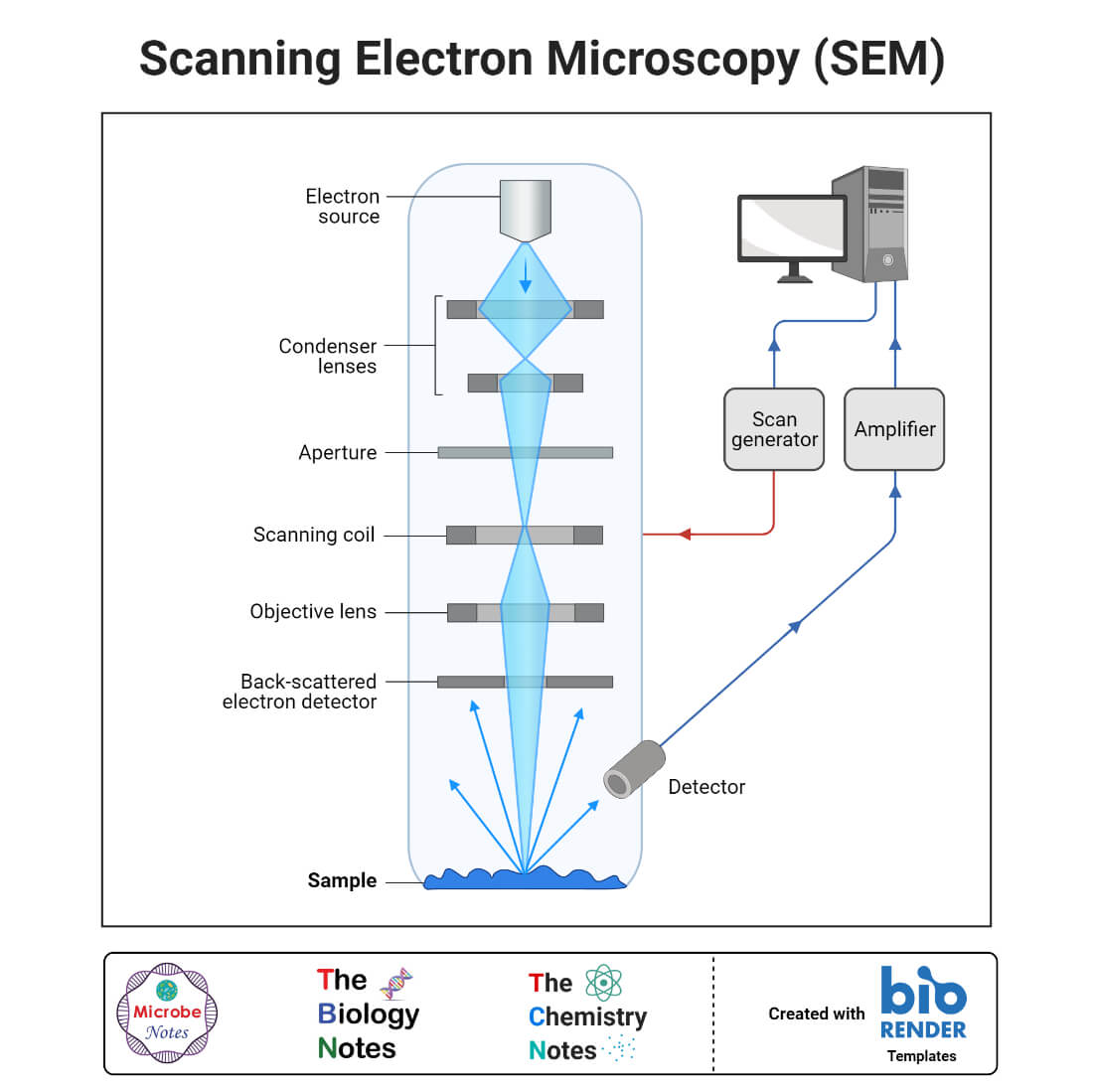
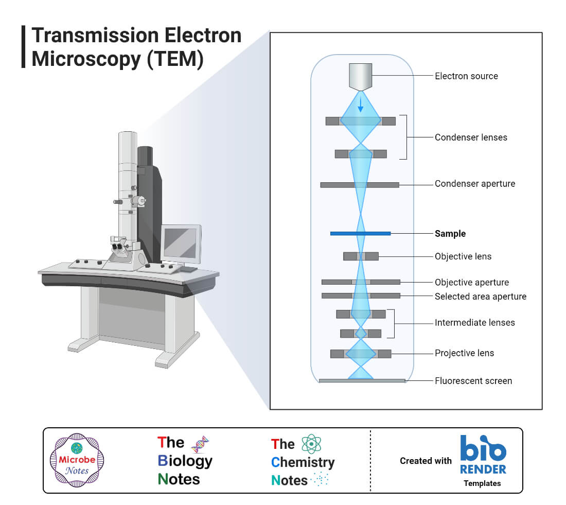
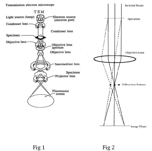
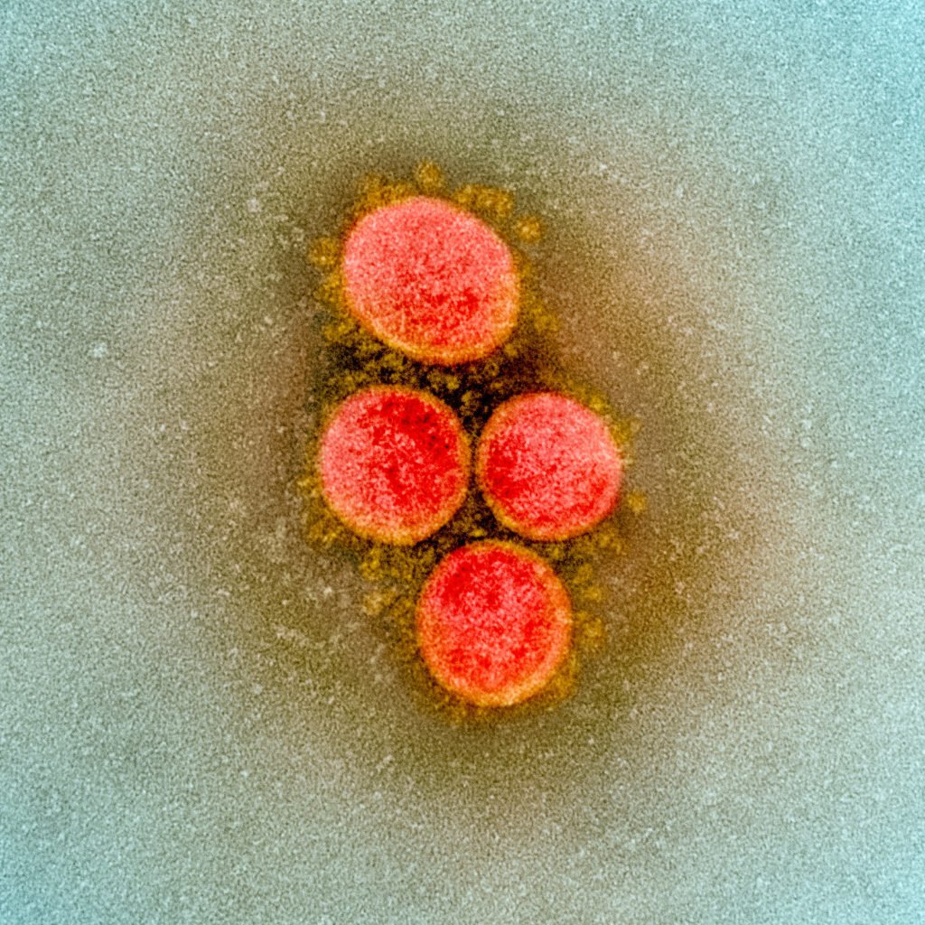
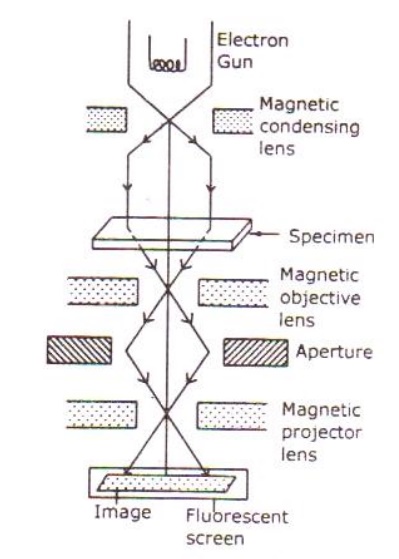
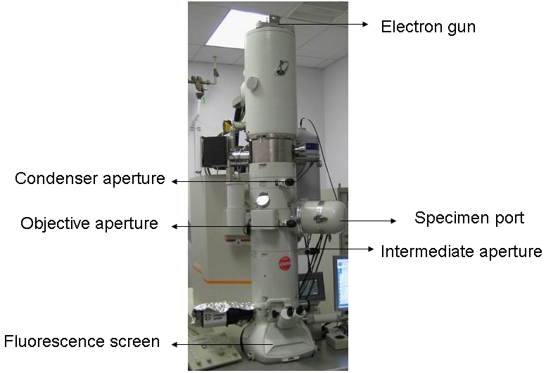
Scanning Electron Microscope (SEM)- Definition, Principle, … Web11/03/2022 · The first Scanning Electron Microscope was initially made by Mafred von Ardenne in 1937 with an aim to surpass the transmission electron Microscope. He used high-resolution power to scan a small raster using a beam of electrons that were focused on the raster. He also aimed at reducing the problems of chromatic aberrations images … (A and B) Electron micrograph of a cell labeled for/5-tubulin followed... Transmission electron microscopy has proven to be a powerful tool in providing information about nanoparticle uptake, biodistribution and relationships with ... Transmission Electron Microscope (TEM)- Definition, Principle, … Web19/05/2022 · Parts of a microscope with functions and labeled diagram; Scanning Electron Microscope (SEM)- Definition, Principle, Parts, Images; Transmission Electron Microscope (TEM) Images. Transmission electron micrograph of SARS-CoV-2 virus particles, isolated from a patient. Image captured and color-enhanced at the NIAID … Transmission Electron Microscopy (TEM) - the University of Warwick Figure 2 shows a simple sketch of the path of a beam of electrons in a TEM from just above the specimen and down the column to the phosphor screen. As the ...
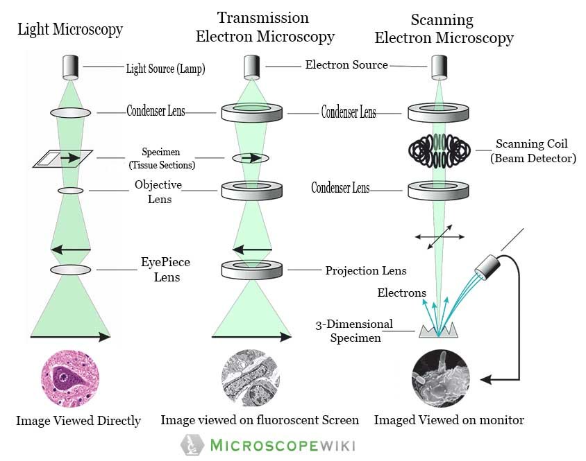
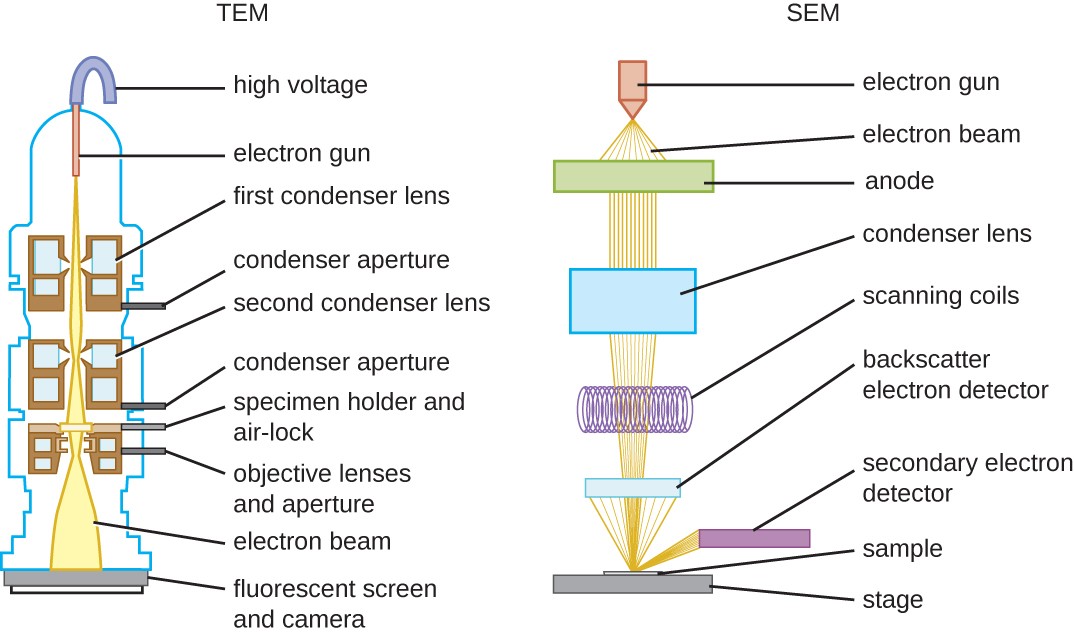
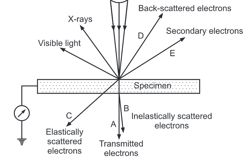
en.wikipedia.org › wiki › Electron_microscopeElectron microscope - Wikipedia An electron microscope is a microscope that uses a beam of accelerated electrons as a source of illumination. As the wavelength of an electron can be up to 100,000 times shorter than that of visible light photons, electron microscopes have a higher resolving power than light microscopes and can reveal the structure of smaller objects. Electron microscope - Wikipedia WebAn electron microscope is a microscope that uses a beam of accelerated electrons as a source of illumination. As the wavelength of an electron can be up to 100,000 times shorter than that of visible light photons, electron microscopes have a higher resolving power than light microscopes and can reveal the structure of smaller objects. A scanning … Transmission Electron Microscopy - an overview | ScienceDirect … WebMiroslaw Jonasz, Georges R. Fournier, in Light Scattering by Particles in Water, 2007. 5.7.8 Transmission electron microscopy (TEM). TEM offers a significantly enhanced resolution (0.0001 μm), about one to two orders of magnitude higher than that of the SEM. However, due to the complex process of sample preparation and time-consuming analysis, this … Fluorescence microscope - Wikipedia WebA fluorescence microscope is an optical microscope that uses fluorescence instead of, or in addition to, scattering, reflection, and attenuation or absorption, to study the properties of organic or inorganic substances. "Fluorescence microscope" refers to any microscope that uses fluorescence to generate an image, whether it is a simple set up like an …
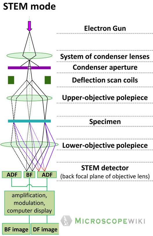
Scanning Transmission Electron Microscopy | SpringerLink
Transmission Electron Microscopy as a Powerful Tool to Investigate ... Despite the high resolution (3–20 nm), imaging is restricted to the surface of the sample. ... Transmission electron microscopy (TEM) provides images obtained by ...

Scanning Electron Microscopy | Central Microscopy Research ...
Transmission electron microscopy DNA sequencing - Wikipedia WebTransmission electron microscopy DNA sequencing is a single-molecule sequencing technology that uses transmission electron microscopy techniques. The method was conceived and developed in the 1960s and 70s, but lost favor when the extent of damage to the sample was recognized. In order for DNA to be clearly visualized under an electron …

Electron Microscope: Principle, Types, Applications – Microbe ...
What Is the Adenovirus? - Healthline Web27/04/2022 · The adenovirus is spread by close personal contact with others, like shaking hands or touching. Coughing, sneezing, or touching an object or surface that has the virus and then touching your mouth ...
Light Microscope vs Electron Microscope- 36 Major Differences
researchgate.net › figure › Low-magnificationLow magnification electron micrograph of a portion of an ... from publication: Electron microscopy of symbiotic bacteria in developing oocytes of the American cockroach, Periplaneta americana | Oocytes and Ovum | ResearchGate, the professional network for ...

14,929 Electron Microscope Images, Stock Photos & Vectors ...
› figure › TEM-micrograph-ofTEM micrograph of longitudinal section of an apoptotic sperm ... Download scientific diagram | TEM micrograph of longitudinal section of an apoptotic sperm characterized by marginated chromatin (mCh) and altered acrosome (aA). A large cytoplasmic residue (CR ...

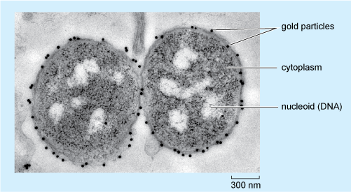
Transmission electron microscopy (TEM). Ten-nanometre gold ...
en.wikipedia.org › wiki › Transmission_electronTransmission electron microscopy DNA sequencing - Wikipedia The DNA molecules must be stretched out on a thin, solid substrate so that order of the labeled bases will be clearly visible on the electron micrograph. Molecular combing is a technique that utilizes the force of a receding air-water interface to extend DNA molecules, leaving them irreversibly bound to a silane layer once dry.
Transmission Electron Microscope: Definition, Parts, Working ...
› figure › A-Electron(A) Electron micrograph of a conventional thin section (∼ 80 ... Download scientific diagram | (A) Electron micrograph of a conventional thin section (∼ 80 nm thickness) shows typical LBs in the cytoplasm of a resting human eosinophil as round, electron-dense ...

Transmission electron micrograph of a thin section through ...
Conventional transmission electron microscopy - PMC - NCBI The most frequently used TEM application in cell biology entails imaging stained thin sections of plastic-embedded cells by passage of an electron beam through ...

Transmission electron micrograph (TEM) showing the nucleus ...
Virtual EM Micrograph List | histology - University of Michigan WebFat Cells: This electron micrograph depicts mature fat cells. You can see one large lipid droplet in the cytoplasm of each cell. The nuclei of many cells are not included in the field of view. Brown fat cells would have several small lipid droplets all of which would be roughly the same size. Remember that each fat cell is enclosed by a thin basal lamina …
Transmission Electron Microscope (TEM)- Definition, Principle ...
The Transmission Electron Microscope | CCBER TEMs employ a high voltage electron beam in order to create an image. An electron gun at the top of a TEM emits electrons that travel through the microscope's ...
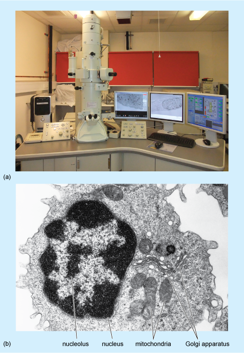
A tour of the cell: View as single page
Transmission electron microscopy - Wikipedia Transmission electron microscopy (TEM) is a microscopy technique in which a beam of electrons is transmitted through a specimen to form an image.
Transmission Electron Microscope (TEM)- Definition, Principle ...
A tour of the cell: View as single page
Transmission electron microscopy - Wikipedia
Electron Microscope- Definition, Principle, Types, Uses ...
Transmission Electron Microscope (TEM)- Definition, Principle ...
Transmission Electron Microscopy (TEM)
Electron Microscope Principle, Uses, Types and Images ...

Transmission electron microscopy - Wikipedia
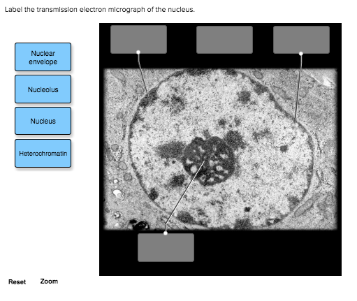
Solved Label the transmission electron micrograph of the ...

anatomy 10.png - Label the transmission electron micrograph ...
Transmission Electron Microscope (TEM)- Definition, Principle ...
Electron Microscope Principle, Uses, Types and Images ...
Transmission electron micrograph (TEM) identifying immunogold ...

Transmission electron microscope (TEM) micrograph showing the ...
Instruments of Microscopy | Microbiology | | Course Hero
8.2: Transmission Electron Microscopy - Chemistry LibreTexts
Preparation of plant cells for transmission electron ...
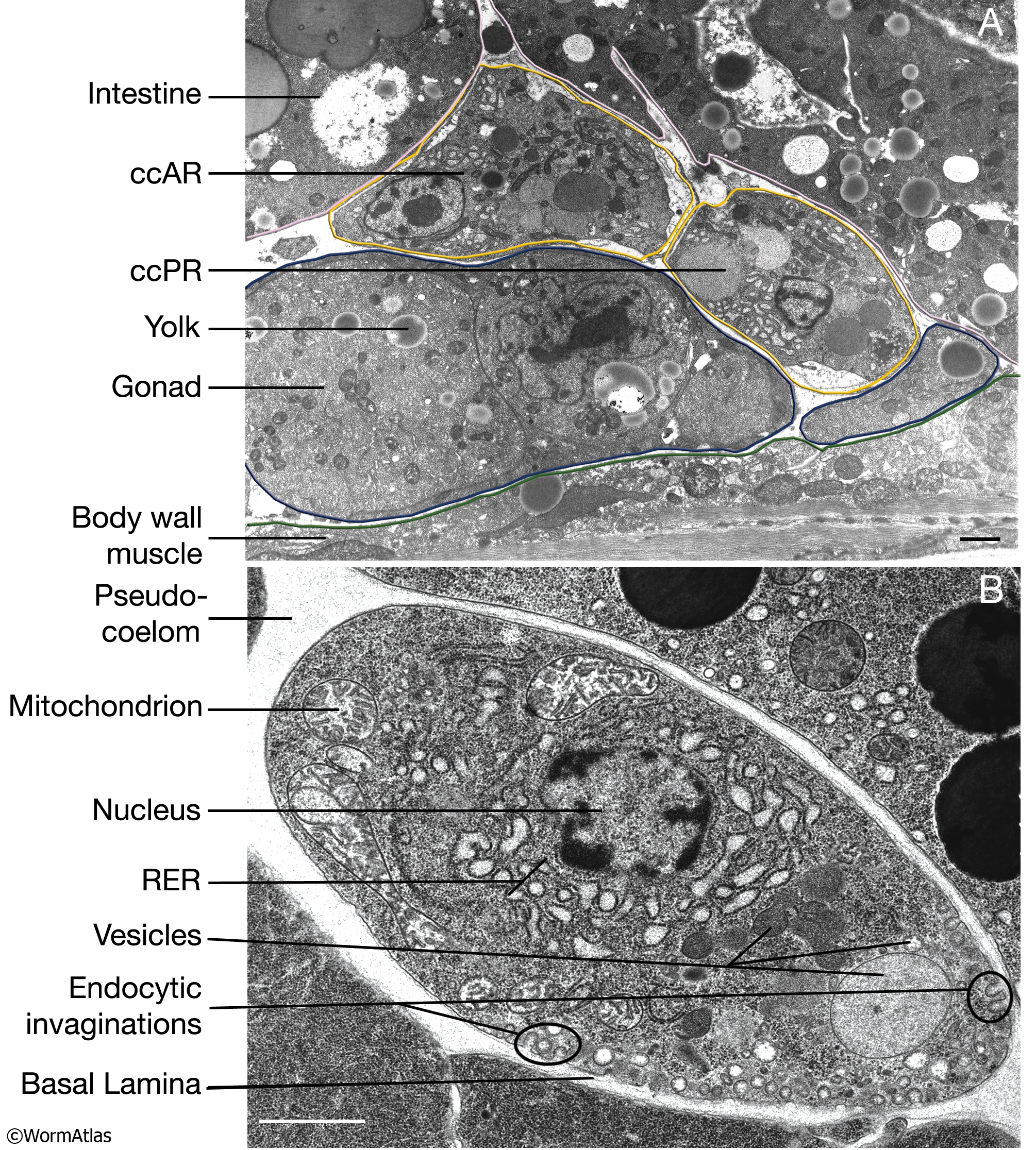
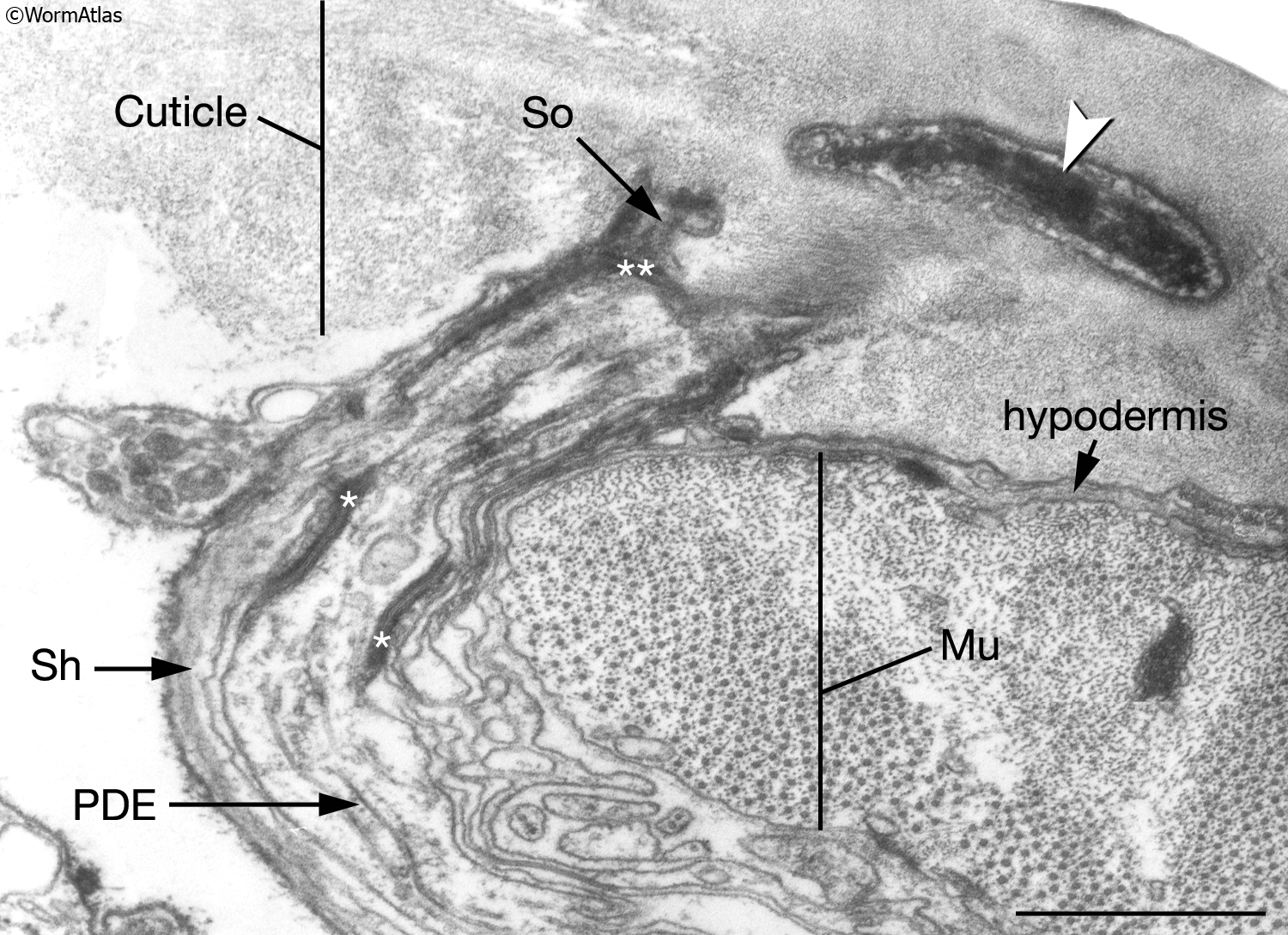
CcFIG 5 Legend

Transmission electron microscopy images of immunogold-labeled ...
Transmission Electron microscope - Principle, Construction ...
IntroFIG 4B Legend
8.2: Transmission Electron Microscopy - Chemistry LibreTexts

Transmission Electron Microscopy of Shape-Controlled ...
Correlative fluorescence microscopy, transmission electron ...

Microscopy

Solved Mitochondrion Nucleus Vesicle Peroxisome | Chegg.com

Transmission electron microscopy - Wikipedia

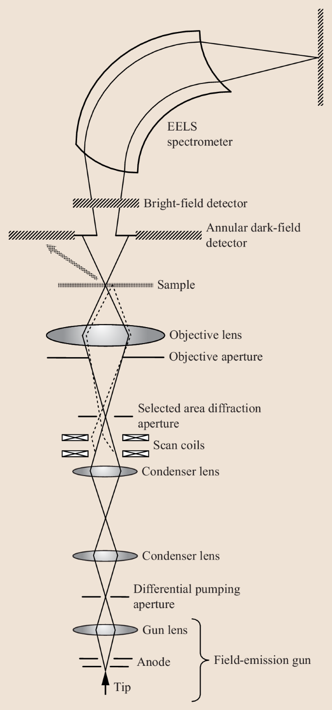
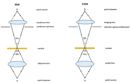
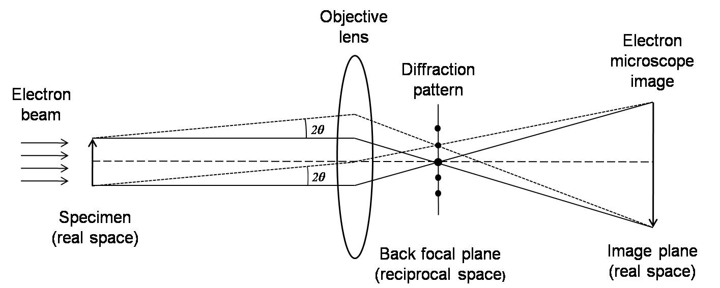
Ray diagrams showing the important optical elements for (a ...

SEM vs TEM | Technology Networks
Transmission electron microscopy cells hi-res stock ...
Transmission Electron Microscope (TEM) - BIOLOGY EASE
Post a Comment for "42 transmission electron micrograph labeled"