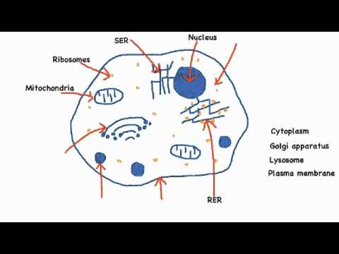
38 draw and label cell
Metaphase in Mitosis and Meiosis (Metaphase 1 and 2) Metaphase Definition. Metaphase is a stage of the cell cycle occurring in both mitosis and meiosis cell division processes. During metaphase in mitosis and meiosis, the chromosomes condense and they become visible and distinguishable during alignment at the center of the dividing cell, to form a metaphase plate at the center of the cell. Drawing Of A Plant Cell With Labels : Animal Cell Diagram High ... Browse draw and label plant cell resources on teachers pay teachers, a marketplace trusted by millions of teachers for original . Plant cells contain large central vacuoles whereas animal cells contain numerous small vacuoles. It determines the function and development of the cell · (b) golgi apparatus: Each label for more information.
Eukaryotic Cell: Structure, Characteristics & Diagram - Embibe Eukaryotic Cell Definition Eukaryotic cell refers to the cell whose genetic material is surrounded by the nuclear membrane, i.e. has a well-defined nucleus and other membrane-bound organelles. "Eu" means true and "karyon" means nucleus. Eukaryotic Cell: Characteristics All living organisms are made of cells.

Draw and label cell
› watchHow to make a Flip Book Animation - YouTube Make a simple animation with a sticky note pad and a pen or pencil. This is a great activity for the young and the old. You will learn how to move things f... › cells › bactcellInteractive Bacteria Cell Model - CELLS alive In the space are enzymes and other proteins that help digest and move nutrients into the cell. Cell Wall: Composed of peptidoglycan (polysaccharides + protein), the cell wall maintains the overall shape of a bacterial cell. The three primary shapes in bacteria are coccus (spherical), bacillus (rod-shaped) and spirillum (spiral). Plant Cell- Definition, Structure, Parts, Functions, Labeled Diagram The cell wall is made up of two layers, a middle lamella, and a primary cell wall and sometimes a secondary cell wall. The middle lamella acts as the strengthening layer between the primary walls of the neighboring cells. The primary wall is made up of cellulose underlying the cells that are dividing and maturing.
Draw and label cell. clinicaltrials.gov › ct2 › showNeoadjuvant Durvalumab Alone or in Combination With Novel ... Jan 07, 2019 · None (Open Label) Primary Purpose: Treatment: Official Title: A Phase 2 Open-label, Multicenter, Randomized, Multidrug Platform Study of Neoadjuvant Durvalumab Alone or in Combination With Novel Agents in Subjects With Resectable, Early-stage (I [> 2 cm] to IIIA) Non-small Cell Lung Cancer (NeoCOAST) Actual Study Start Date : March 8, 2019 Well Labelled Diagram Of Animal Cell - A Draw A Well Labeled Diagram Of ... Beranda / Well Labelled Diagram Of Animal Cell - A Draw A Well Labeled Diagram Of Animal Cell B Name The Organelle Which Is Found Only In Animal Cells What Are Its Functions Biology Topperlearning Com 72sonq166 - (a) ... provide the labels for the structures involved in the reflex act when a person steps on a tack and jerks their leg away. The ... Plant and Animal Cell: Labeled Diagram, Structure, Function - EMBIBE Plant Cell: Plant cells are eukaryotic cells with a true nucleus along with specialized structures called organelles that carry out certain specific functions. Animal Cell: An animal cell is a type of eukaryotic cell that lacks a cell wall and has a true, membrane-bound nucleus along with other cellular organelles. Cell Cycle: Phases, Diagram, Stage, and Checkpoints Gap 2 / G2 Phase. o During the gap between DNA synthesis and mitosis, the cell will continue to grow. o Cellular organelles continue to duplicate. o RNA and protein (especially tubulin for microtubules) are actively synthesized. o The G2 checkpoint control mechanism ensures that everything is ready to enter the M (mitosis/meiosis) phase and divide.
› cell-membrane-373364Cell Membrane Function and Structure - ThoughtCo Oct 07, 2019 · Microscopic view of phospholipids. Stocktrek Images / Getty Images. Phospholipids are a major component of cell membranes.Phospholipids form a lipid bilayer in which their hydrophilic (attracted to water) head areas spontaneously arrange to face the aqueous cytosol and the extracellular fluid, while their hydrophobic (repelled by water) tail areas face away from the cytosol and extracellular ... How to: Custom Draw Cells Depending Upon Cell Values The InitButtonEdit method configures the Name column's editor as follows: Sets the Kind property to ButtonPredefines.Glyph to display both the image and caption within the button. Sets the editor's TextEditStyle property to TextEditStyles.HideTextEditor. This hides the text box and stretches the editor's button to fit a cell. How to Draw a Custom Table in Microsoft Word To draw your table, go to the Insert tab and click the Table drop-down arrow. Select "Draw Table." You'll see your cursor change to a pencil icon. Drag to draw the outline of the table first. You can make it any size you need and use a square or a rectangle. Next, draw the columns, rows, or individual cells. Shape.Cells property (Visio) | Microsoft Docs where xxx is one of these cells: Label, Prompt, SortKey, Type, Format, Invisible, ... Use the Cells property to get a Cell object by using the cell's local name. ... End If 'Draw a rectangle on the active page. Set vsoShape = vsoPage.DrawRectangle(1, 5, 5, 1) 'Add a scratch section and add a row to the scratch section. ...
(Get Answer) - 3. Draw and label an animal and plant cell ... - Transtutors Draw and label an animal and plant cell (eukaryotic cell). Label and give a function for these organelles/structures and give a function(s) for each: Cell (plasma) membrane Cytoplasm Cell wall Ribosome Nucleus/nuclear membrane/nucleolus Rough... Different Size, Shape and Arrangement of Bacterial Cells Size of Bacterial Cell. The average diameter of spherical bacteria is 0.5-2.0 µm. For rod-shaped or filamentous bacteria, length is 1-10 µm and diameter is 0.25-1 .0 µm. E. coli , a bacillus of about average size is 1.1 to 1.5 µm wide by 2.0 to 6.0 µm long. Spirochaetes occasionally reach 500 µm in length and the cyanobacterium. How to Draw a Circuit Diagram - Edraw - Edrawsoft EdrawMax is an advanced all-in-one diagramming tool for creating professional flowcharts, org charts, mind maps, network diagrams, UML diagrams, floor plans, electrical diagrams, science illustrations, and more. Just try it, you will love it! Free Download Buy Now › cells › 3dcellInteractive Cell Models - CELLS alive Living cells are divided into two types - prokaryotic and eukaryotic (sometimes spelled procaryotic and eucaryotic). This division is based on internal complexity. The following interactive animations provide graphic roadmaps to the organization of both of these cell types.
15 Cord Management Life Hacks for No More Tangled Wires
Cells Diagram | Science Illustration Solutions - Edrawsoft Edraw software offers you lots of symbols used in cells diagram like cell structure, paramecium, squamous cell, cell division, bacteria, cell membrane, eggs, sperm, zygote, an animal cell, SARS, tobacco mosaic, adenovirus, coliphage, herpesvirus, AIDS, pollen, plant cell model, onion tissue, etc. Cells Diagram Examples

Mitosis And Meiosis Drawing Worksheet - Blogger Mitosis Meiosis Drawing Worksheet. Discuss cell changes during interphase. Draw and label the four stages of mitosis. Meiosis Worksheet - Mr. For Students 7th - 12th. A basic worksheet that walks budding biologists through observing drawing and describing cells in different stages of mitosis. Describe and explain the various stages of cell ...
Draw a diagram of human sperm. Label only those parts along with their ...
Interphase- Definition, Stages, Cell cycle, Diagram, Video The synthesis (S) phase is the phase of cell copying or cell duplication of its DNA of its entire genome. Gap 1 (G1) This is the phase in which the cell undergoes normal growth and cell function synthesizing high amounts of proteins. The cell increases in size and volume as more cell organelles are produced.
Biology diagram worksheet
TUTORIAL CHEMISTRY SK025 - Flip eBook Pages 1-50 | AnyFlip A galvanic cell consists of an Al electrode in 1.00 M Al(NO3)3, a Pb electrode in 1.00 M Pb(NO3)2 and a KCl salt bridge. i. Draw and label the cell diagram. ii. Write cell notation of the reaction. iii. Write the overall cell reaction. iv. Calculate the standard cell potential. v. Which electrode will increase in weight? Explain your answer.

V Ling: 01.11
Draw Animal Cell And Plant Cell With Labelling / Draw An Animal Cell ... Browse draw and label plant cell resources on teachers pay teachers,. Draw a out line of animal cell, put lot of bends as shown to represent flexible plasma membrane. Help kids learn, recall, and apply the basic differences between plant and animal cells with our . The animal cell includes 17 organelles, and the plant cell .

V Ling: 01.11
Sneak Peek - AP Drawing - Acellus Student Blog Label. 354 Comments . most voted. newest oldest. Inline Feedbacks. View all comments. kitty. August 5, 2021 8:28 am ... Drawing is the best! it makes me so happy! Like if it makes you feel happy to! 48. Reply. juice WRLD August 9, 2021 7:33 am wow cool. 49. Reply. rumaysah
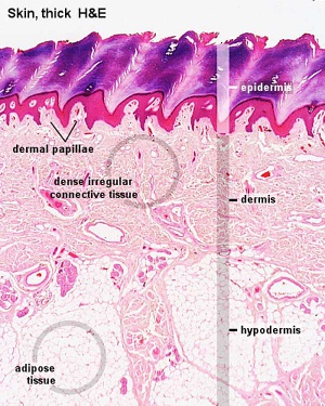
ANAT2241 Connective Tissue Components - Embryology
Shapes.AddLabel method (Excel) | Microsoft Docs Creates a label. Returns a Shape object that represents the new label. Syntax expression. AddLabel ( Orientation, Left, Top, Width, Height) expression A variable that represents a Shapes object. Parameters Return value Shape Example This example adds a vertical label that contains the text Test Label to myDocument. VB
2.3.1 Draw and label a diagram of the ultrastructure of a liver cell as ...
Learn the parts of a cell with diagrams and cell quizzes For this exercise we'll start with an image of a cell diagram ready labeled. Study this and make sure that you're clear about which structure is found where. Cell diagram unlabeled It's time to label the cell yourself! As you fill in the cell structure worksheet, remember the functions of each part of the cell that you learned in the video.

Plant cell model - YouTube
Drawing Of A Plant Cell With Labelled Parts - Structure Of Animal And ... Draw a plant cell and label the parts. Hi friends,plant cell, the basic unit of all plants. The chloroplast is present only in plant cells. It determines the function and development of the cell · (b) golgi apparatus: Draw a labelled diagram of a plant . It packages materials coming from the endoplasmic .
Post a Comment for "38 draw and label cell"